


Clean and in beautiful condition. Unit was pulled from premiere state facility during a facility upgrade. This system was regularly maintained and the department reported the unit fully functional. In our onsite tests, both transponders scanned cleanly. Interface operations functioned perfectly and the monitor was clear and brought. The system as a whole was well behaved and quiet.
This system has the following active licensed options installed:
- Vivid 7 Dimensions
- Blood Flow Imaging
- EchoPAC
- EchoStress
- MPEGvue
- Ditcom Media
- Anatomical M Mode
- Tissue Velocity Imaging & Tissue Tracking
- TrackingM3S/M4S Support & True Speed
Comes as shown with M4S and 10S transducer probes, system backup thumb drive, ECG cables and accessories as shown in photos.
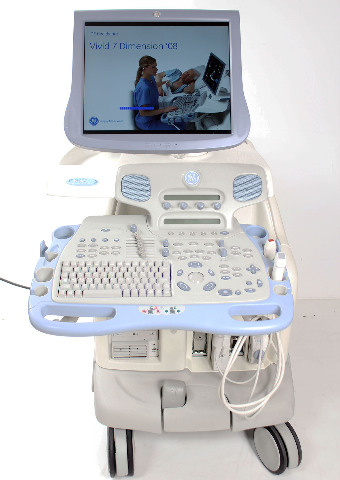
GE Vivid 7 Dimension Ultrasound
The GE Vivid 7 Dimension, with its 2008 release, was a high-performance console ultrasound system that significantly advanced cardiovascular imaging. It was designed to integrate multi-dimensional and 4D capabilities into a standard workflow, offering a new level of diagnostic precision and efficiency. The system was built on GE's TruScan architecture, allowing for raw data storage and advanced post-processing, making it a powerful tool for cardiac, vascular, and shared-service applications.
Key Features
-
Real-Time 4D Imaging: The Vivid 7 Dimension was a leader in real-time 4D imaging, allowing for online, ungated, and unspliced full-volume acquisitions of the beating heart. This provided a comprehensive view of cardiac anatomy and function.
-
TruScan Architecture: This core technology digitally acquires and stores ultrasound data in a raw state, preserving image integrity. This enables post-exam measurement, optimization, and analysis without losing original image quality.
-
Multi-Dimensional Imaging: The system introduced tri-plane imaging, a first in cardiac ultrasound, for the simultaneous display of three images from the same heartbeat. This enhanced the accuracy of assessments in grayscale, color, and Tissue Velocity Imaging (TVI).
-
Advanced Quantitative Tools: Features like Automated Function Imaging (AFI) and Advanced Tissue Synchronization Imaging (TSI) allowed for the assessment and quantification of left ventricle wall motion and synchronicity, crucial for heart failure and CRT patients.
-
Ultra-Definition Imaging: New image processing filters, including Ultra-Definition Clarity Control, helped reduce noise while maintaining fine detail, leading to improved image quality for various applications.
-
B-Flow Imaging: This digital imaging technique provided real-time visualization of blood flow without the typical "bleeding" of color Doppler, offering better delineation of vessel walls.
-
Streamlined Workflow: The system integrated a flexible reporting package with structured findings, programmable user presets, and a digital clipboard, all designed to increase clinical productivity and speed up exam times.
-
DICOM Connectivity: Full DICOM 3.0 compliance for seamless integration into hospital networks (PACS), enabling remote image monitoring and data sharing.
Specifications
-
System Type: Console Ultrasound
-
Monitor: 17" or 21" High-Resolution LCD monitor with tilt and swivel
-
Probe Ports: 3 active probe ports
-
Imaging Modes:
-
2D (B-Mode)
-
M-Mode & Anatomical M-Mode
-
Color Doppler & Power Doppler
-
Pulsed Wave (PW) Doppler
-
Continuous Wave (CW) Doppler
-
Tissue Doppler Imaging (TDI)
-
B-Flow
-
Real-Time 4D
-
-
Dimensions:
-
Height: 54.1 in (137.5 cm)
-
Width: 25.2 in (64.0 cm)
-
Depth: 35.4 in (90.0 cm)
-
-
Weight: Approximately 403 lbs (183 kg) with monitor, without peripherals
-
Digital Storage: Internal hard drive for patient data, loops, and images.
-
Connectivity:
-
DICOM 3.0 (Storage, Worklist, Print)
-
USB 2.0
-
LAN Ethernet
-
-
Applications:
-
Adult & Pediatric Cardiac
-
Vascular
-
Abdominal
-
Small Parts (Breast, Thyroid)
-
Obstetrics/Gynecology
-
Urology
-
-
Advanced Options (Often Included):
-
4D Volume & 4D Volume Color Flow
-
Automated Function Imaging (AFI)
-
Advanced Tissue Synchronization Imaging (TSI)
-
Strain Rate Imaging (SRI)
-
Blood Flow Imaging (BFI)
-
Contrast Imaging
-
EchoPAC
-
EchoStress
-